
Bone Pain in Seniors: How to Choose Between MRI, CT, and DEXA Scans
Bone pain is common in older adults, but it should not always be considered a normal part of ageing. Persistent back, hip, knee, or other bone pain, especially after a minor fall or without an obvious injury, may be a sign of osteoporosis, a hidden fracture, arthritis, or another bone condition that needs proper evaluation.
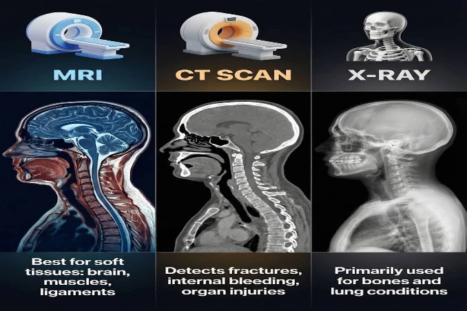
There is no single “best” scan for every type of bone pain. MRI is often recommended for hidden fractures, spine problems, and soft tissue injuries; CT Scan provides detailed images of bone structure and complex fractures; while DEXA Scan is the gold standard for measuring bone density and diagnosing osteoporosis. The right scan depends on the underlying cause of the pain and your doctor’s assessment.
At Orbit Imaging, we provide advanced MRI, CT Scan, and DEXA Scan services to help diagnose bone-related conditions accurately. With over 21 years of experience, expert radiologists, and conveniently located centres across Delhi NCR, we are committed to delivering reliable imaging that supports timely diagnosis and treatment.
Why Bone Pain Should Never Be Ignored After 60
Bone pain is not a normal part of ageing. If it lasts for more than 2–3 weeks, starts after a minor fall, or affects your daily activities, it should be evaluated. It may be caused by osteoporosis, a hidden fracture, arthritis, or another bone condition.
Consult a doctor if you have:
- Bone pain lasting more than 2–3 weeks
- Pain after a minor fall or injury
- Sudden back or hip pain
- Pain that wakes you at night
- Difficulty standing or walking
- Repeated fractures
- Gradual loss of height
Early imaging helps identify the real cause of the pain and allows treatment to begin before the condition worsens.
The First Question Isn't "Which Scan?" It's "What Kind of Bone Pain Do You Have?"
|
What You Are Feeling |
Possible Cause |
Recommended Scan |
|
Sudden back pain after lifting something |
Compression fracture |
MRI |
|
Hip pain after a minor fall |
Hidden hip fracture |
MRI or CT Scan |
|
Knee pain with swelling |
Arthritis or ligament injury |
MRI |
|
Persistent deep bone pain |
Bone infection or bone damage |
MRI |
|
Repeated fractures |
Osteoporosis |
DEXA Scan |
|
Gradual loss of height |
Bone loss |
DEXA Scan |
|
Complex fracture |
Multiple bone fragments |
CT Scan |
Every scan has a different purpose. MRI is commonly used to detect hidden fractures and soft tissue injuries; a CT Scan provides detailed images of bone structure and complex fractures, while a DEXA Scan measures bone strength and helps diagnose osteoporosis.
In some cases, doctors may recommend more than one imaging test. For example, an MRI may identify the cause of persistent back pain, while a DEXA scan checks whether osteoporosis has weakened the bones. The right scan depends on your condition, which is why it is always chosen after a medical evaluation.
MRI vs CT Scan vs DEXA Scan: Which One Is Right?
The three imaging tests are often mentioned together, but they serve very different purposes. Understanding what each scan actually looks for can make conversations with your doctor much easier.
The table below provides a quick comparison.
|
Feature |
MRI Scan |
CT Scan |
DEXA Scan |
|
Primary purpose |
Detect hidden injuries and soft tissue problems |
Detailed bone structure |
Measure bone density |
|
Best for |
Hidden fractures, spine problems, bone marrow, ligaments, infections |
Complex fractures, joint injuries, surgical planning |
Osteoporosis and fracture risk |
|
Not ideal for |
Measuring bone strength |
Measuring bone density |
Finding the cause of active bone pain |
|
Radiation |
None |
Uses X-rays |
Very low radiation |
|
Typical duration |
Around 20–45 minutes |
Usually 5–15 minutes |
Around 10–20 minutes |
|
Doctors commonly recommend when |
Pain source is unclear, or hidden injury is suspected |
Detailed bone assessment is needed |
Osteoporosis is suspected, or fracture risk needs assessment |
Although these scans may seem similar to patients, they answer completely different questions.
Think of it this way:
- MRI looks for what is happening inside the bone and surrounding tissues.
- CT Scan shows the shape and structure of the bone in exceptional detail.
- DEXA Scan measures how strong or weak the bones are.
Because they serve different purposes, one scan cannot simply replace another.
MRI Scan: Best for Finding Hidden Bone and Soft Tissue Problems
MRI (Magnetic Resonance Imaging) uses magnets and radio waves to create detailed images without using radiation. It is often recommended when bone pain continues but the cause is not clearly visible on an X-ray.
MRI is commonly used to detect:
- Hidden or hairline fractures
- Spine and hip problems
- Bone marrow changes
- Ligament and tendon injuries
- Joint cartilage damage
- Bone infections
- Nerve compression
One of MRI’s biggest advantages is that it can detect early bone marrow changes, helping identify hidden fractures before they become visible on other imaging tests. It also provides clear images of muscles, ligaments, and nerves, making it useful when pain may not be coming directly from the bone.
CT Scan: Best for Detailed Bone Structure and Complex Fractures
- A CT (Computed Tomography) Scan uses multiple X-ray images to create detailed cross-sectional and 3D views of bones. It is commonly recommended when doctors need a clear view of a fracture or bone injury.
CT scans are commonly used for:
- Complex or multiple fractures
- Pelvic and joint injuries
- Facial bone fractures
- Surgical planning
- Checking fracture healing
- Assessing bone alignment
CT scans provide highly detailed images of the bone’s structure, helping doctors understand the severity of an injury and plan the most appropriate treatment. They are also much faster than MRI, making them useful in emergencies. If a contrast CT scan is needed, your doctor may recommend a kidney function test beforehand, especially for older adults.
DEXA Scan: The Gold Standard for Measuring Bone Strength
A DEXA (Dual-Energy X-ray Absorptiometry) scan is very different from MRI and CT.
Instead of searching for the cause of pain, it measures bone mineral density, allowing doctors to assess how strong or weak the bones are.
DEXA scans are commonly used to:
- Diagnose osteoporosis
- Detect osteopenia (early bone loss)
- Estimate future fracture risk
- Monitor response to osteoporosis treatment
- Assess long-term bone health
The scan is quick, painless, and uses extremely low levels of radiation.
One important point that many people misunderstand is that a DEXA scan does not identify why a particular bone hurts.
For example, if an elderly patient has severe hip pain after slipping in the bathroom, a DEXA scan cannot confirm whether a fracture is present. It only measures bone density.
Likewise, an MRI cannot measure bone density with the accuracy needed to diagnose osteoporosis.
Because of this difference, doctors sometimes recommend both scans.
For instance, a patient with persistent back pain may undergo:
- An MRI to identify whether a spinal compression fracture is causing today’s pain.
- A DEXA scan to determine whether osteoporosis contributed to the fracture and whether long-term treatment is needed.
Understanding this distinction helps patients avoid one of the most common misconceptions—that a single scan answers every question.
Can MRI Detect Osteoporosis?
This is one of the most common questions seniors ask.
The short answer is no.
MRI can detect complications caused by osteoporosis, such as compression fractures or bone marrow swelling, but it cannot measure bone mineral density, which is essential for diagnosing osteoporosis.
That is why doctors continue to rely on DEXA scans, which remain the internationally accepted standard for measuring bone strength and estimating fracture risk.
Think of it this way:
- MRI answers: Why is this bone hurting today?
- DEXA answers: How strong are these bones overall?
The two tests complement each other rather than compete with each other.
Can a CT Scan Replace a DEXA Scan?
Again, the answer is no.
CT scans create extremely detailed images of bone structure, making them excellent for diagnosing fractures and understanding complex injuries. However, they are not designed to measure bone mineral density with the accuracy required for osteoporosis diagnosis.
If osteoporosis is suspected, doctors generally recommend a dedicated DEXA scan instead of relying on CT findings.
Understanding this difference helps patients avoid unnecessary investigations and ensures they receive the imaging test that best matches their condition.
When Should Bone Pain Be Investigated Immediately?
Not every ache requires emergency imaging. Mild discomfort after physical activity often improves with rest and appropriate care.
However, certain symptoms should never be ignored because they may indicate a fracture, serious infection, or another condition that requires urgent evaluation.
Seek medical attention promptly if you experience:
- Severe pain after even a minor fall, especially around the hip or spine.
- Inability to stand or bear weight on one leg.
- Sudden back pain after lifting something light.
- Persistent pain that continues for several weeks despite rest.
- Bone pain accompanied by fever, redness, or swelling.
- Pain that repeatedly wakes you during the night.
- Sudden weakness, numbness, or difficulty controlling bowel or bladder function along with severe back pain. This requires immediate emergency evaluation.
- Unexplained weight loss together with persistent bone pain, which needs further medical assessment.
Early diagnosis often means earlier treatment, fewer complications, and a better recovery.
Why Self-Diagnosing Bone Pain Can Delay Treatment
Many seniors ignore bone pain or try to manage it at home, which can delay the right diagnosis. Common mistakes include:
- Assuming the pain is just arthritis or old age
- Relying on painkillers or pain relief gels
- Ignoring pain after a minor fall
- Waiting for the pain to improve on its own
In older adults, even a small slip or lifting a light object can cause a hidden fracture due to weak bones. Imaging tests help detect these problems early, allowing treatment to begin before the condition becomes more serious.
How Doctors Decide Between MRI, CT Scan, and DEXA
Patients often wonder why two people with similar symptoms receive different scan recommendations.
The answer is that imaging decisions are based on clinical evaluation rather than a fixed rule.
Doctors commonly consider questions such as:
- Did the pain begin after a fall or injury?
- Is the pain sudden or gradually worsening?
- Is movement restricted?
- Is osteoporosis suspected?
- Has the patient experienced repeated fractures?
- Is surgery being considered?
- Does the pain seem to originate from the bone, joint, muscles, or nerves?
- Have previous X-rays already been performed?
These answers help determine which imaging test is likely to provide the clearest diagnostic information.
In some cases, imaging may begin with one test and later be followed by another if additional information is needed.
Preparing for Your Imaging Test
Most imaging tests require very little preparation, but following your doctor’s instructions helps ensure accurate results.
Before an MRI
- Remove jewellery, watches, hearing aids, and other metal objects.
- Inform the radiology team if you have a pacemaker, aneurysm clips, cochlear implant, or other implanted medical devices.
- Wear comfortable clothing or use the gown provided.
- If an MRI with contrast is advised, your doctor may recommend checking kidney function beforehand.
Before a CT Scan
- Wear comfortable clothing.
- Follow any fasting instructions if contrast material is being used.
- Tell your healthcare provider about kidney disease, allergies, or previous reactions to contrast dye.
- Blood tests such as serum creatinine may be required before contrast-enhanced CT scans in some elderly patients.
Before a DEXA Scan
- Wear clothing without heavy metal zippers or buttons if possible.
- Follow your doctor’s advice regarding calcium supplements before the test.
- The scan is quick, painless, and usually completed within a few minutes.
If you are unsure about preparation, our team at Orbit Imaging is always available to guide you before your appointment.
Why Choose Orbit Imaging for Bone Imaging in Delhi NCR?
Choosing the right scan is important, but so is choosing the right imaging centre. Clear images and accurate reporting help doctors diagnose bone problems with greater confidence.
At Orbit Imaging, we have been providing trusted diagnostic imaging for 21+ years with advanced technology and experienced radiologists.
Why choose Orbit Imaging?
- Advanced MRI, CT Scan, and DEXA Scan technology
- Experienced MD & MBBS radiologists
- High-resolution, accurate imaging
- 1 Crore+ patients served
- Trusted by doctors across Delhi NCR
- Affordable pricing
- Fast appointments and timely reports
- Senior-friendly care and support
Whether your doctor recommends an MRI, CT Scan, or DEXA Scan, our experienced team helps deliver accurate imaging for faster diagnosis and better treatment planning.
Visit Your Nearest Orbit Imaging Centre
Travelling long distances can be uncomfortable for elderly patients dealing with bone or joint pain. That is why Orbit Imaging has multiple conveniently located centres across Delhi NCR, allowing families to choose the branch closest to them.
Our active locations include:
- Karol Bagh – Central Delhi, Pusa Road area
- Rohini – Prashant Vihar, Outer Ring Road
- Green Park – Yusuf Sarai, South Delhi
- Dwarka – Sector 7, near Ramphal Chowk
- Dilshad Garden – Main Road, East Delhi
- Gurugram – Sector 52, near Artemis Hospital
Whether you need an MRI, CT Scan, or DEXA Scan, our team is committed to providing accurate imaging with patient comfort and convenience in mind.
Conclusion
Bone pain is not a normal part of ageing and should never be ignored. The right scan depends on the cause—MRI helps detect hidden fractures and soft tissue problems, CT Scan provides detailed bone images, and DEXA Scan measures bone strength to diagnose osteoporosis.
At Orbit Imaging, we offer advanced MRI, CT Scan, and DEXA Scan services with 21+ years of experience, expert radiologists, modern technology, and multiple centres across Delhi NCR to ensure accurate diagnosis and timely reports.
Book your appointment at your nearest Orbit Imaging centre today and get the right scan for the right diagnosis.

4.2 Stars on Google Maps (900+ Reviews)
Free Consultation for your test
Reliable and Affordable diagnostics
NABL, NABH, and QCI-accredited
Crisp Imaging & Justified Pricing
22 Years of Trust & Care
Fill the form & leave it on us!
22+
Years in Diagnostics
1 Cr+
Patients Served
450+
Tests & Scans Available
200+
Radiologists & Technicians
Orbit Imaging & Pathlab–Rohini
Address: First Floor, A-1/5, A Block, Prashant Vihar, Sector 14, Rohini, Delhi, 110085, M: 9289289216
Why Choose Orbit Imaging!
22 Years of Trust & Care
MD & MBBS Radiologists
Lowest Rates in Delhi NCR
Recommended by Leading Doctors
1 Cr+ Patients Served
Cardiology Tests at Orbit Imaging
ECHO
ECG
CT ANGIOGRAPHY
CARDIAC MRI
Radiology Tests at Orbit Imaging
MRI
CT SCAN
ULTRASOUND
PET CT SCAN
MAMMOGRAPHY
BONE DEXA
PFT
Pathology Tests at Orbit Imaging
Neurology Tests at Orbit Imaging
Orbit Imaging & Pathlab
Best Diagnostic Lab with Affordable Rates in Rohini