Introduction
Welcome to a comprehensive exploration of the MRI hip joint process and diagnosis. In this detailed guide, we delve into the intricacies of magnetic resonance imaging (MRI) procedures focused on the hip joint. Our goal is to provide you with an insightful understanding of the diagnostic capabilities of MRI, shedding light on the nuances that set it apart as a crucial tool in assessing hip joint health.
Understanding the Basics of MRI
Magnetic Resonance Imaging Explained
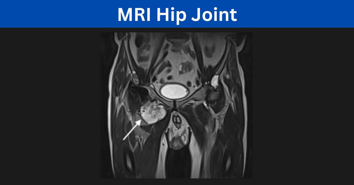
Magnetic Resonance Imaging, commonly known as MRI, is a sophisticated medical imaging technique that utilizes powerful magnets and radio waves to create detailed cross-sectional images of the internal structures of the body. When applied to the hip joint, MRI becomes an invaluable diagnostic tool, offering unparalleled clarity in visualizing soft tissues, bones, and joint structures.
Significance of MRI in Hip Joint Assessment
The hip joint, being a complex structure, requires precise imaging for accurate diagnosis. MRI excels in capturing high-resolution images of the hip joint, enabling healthcare professionals to assess conditions such as labral tears, cartilage damage, and ligament injuries with exceptional accuracy.
The MRI Hip Joint Process
Preparing for the MRI
Before embarking on an MRI journey, proper preparation is essential. Patients are advised to remove any metallic objects and inform the radiologist about existing medical conditions or implants. This ensures a smooth and safe imaging process.
Patient Positioning
Achieving optimal imaging results involves precise patient positioning. For hip joint imaging, patients are typically positioned lying on their back, with the affected hip centered in the MRI machine. The need for stillness during the scan is emphasized to avoid motion artifacts that could compromise image quality.
Contrast Enhancement
In certain cases, contrast agents may be administered to enhance the visibility of specific structures within the hip joint. This is particularly useful in highlighting abnormalities and aiding in the accurate diagnosis of conditions such as hip impingement or synovitis.
Diagnosis Precision with MRI
Detecting Labral Tears
One of the primary benefits of MRI in hip joint diagnosis is its ability to detect labral tears. The labrum is a cartilage structure that surrounds the hip socket, and tears can lead to pain and discomfort. MRI enables clinicians to visualize these tears and determine their extent, guiding treatment decisions effectively.
Evaluating Cartilage Health
MRI’s detailed imaging capabilities extend to the assessment of cartilage health in the hip joint. It allows for the identification of cartilage defects and degeneration, providing vital information for orthopedic specialists in formulating appropriate treatment plans.
Unraveling Ligament Injuries
Ligament injuries in the hip joint, though less common, can significantly impact mobility. MRI plays a pivotal role in unraveling these injuries by capturing detailed images of ligament structures. This aids orthopedic surgeons in devising surgical interventions or rehabilitation strategies.
Advantages of MRI Over Alternative Imaging Techniques
Superior Soft Tissue Resolution
MRI’s superiority lies in its exceptional soft tissue resolution. Unlike traditional X-rays or CT scans, MRI excels in visualizing soft tissues, making it indispensable in the assessment of muscles, tendons, and ligaments surrounding the hip joint.
Non-Invasiveness and Safety
Unlike invasive procedures such as arthroscopy, MRI is a non-invasive imaging technique that poses minimal risk to patients. It eliminates the need for surgical exploration, allowing for a safer and more patient-friendly diagnostic approach.
Conclusion
In conclusion, the MRI hip joint process and diagnosis stand as a pinnacle in the realm of orthopedic diagnostics. Its unparalleled ability to provide detailed, non-invasive images of the hip joint sets it apart as a crucial tool for healthcare professionals. Whether unraveling the complexities of labral tears, assessing cartilage health, or detecting ligament injuries, MRI emerges as the gold standard, guiding precise diagnoses and informed treatment decisions.