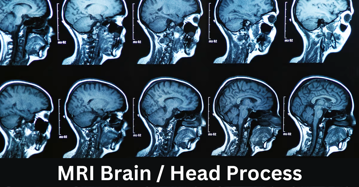
Introduction to MRI Brain/Head Imaging
In the realm of medical diagnostics, Magnetic Resonance Imaging (MRI) has emerged as a revolutionary tool for examining the brain and head without resorting to invasive procedures. The significance of non-ionizing radiation in MRI cannot be overstated, making it a preferred choice for clinicians and patients alike.
Understanding the Basics of MRI Technology
Magnetic Resonance and Radiofrequency Signals
At the core of MRI lies the interaction between magnetic fields and radiofrequency signals. This synergy allows for the creation of detailed images, providing valuable insights into the structure and functioning of the brain.
Role of Strong Magnetic Fields
MRI machines employ strong magnetic fields to align hydrogen atoms within the body temporarily. The subsequent release of energy when these atoms return to their normal state is harnessed to generate precise images.
Step-by-Step Process of MRI Brain/Head Imaging
Patient Preparation
Before the scan, patients are adequately prepared, ensuring optimal results. This includes removing metal objects, wearing appropriate clothing, and addressing any concerns related to claustrophobia.
Coil Placement and Positioning
Coils, strategically placed around the head, enhance the accuracy of imaging. Proper positioning is critical to capturing detailed pictures of the brain’s intricate structures.
Image Acquisition
The MRI machine captures multiple cross-sectional images, which are then compiled to create a comprehensive 3D representation of the brain. The duration of the scan varies but typically ranges from 30 to 60 minutes.
Types of MRI Sequences for Brain/Head Imaging
T1-weighted Images
T1-weighted images highlight the anatomy of the brain, offering clarity on structures like gray matter. These are valuable for detecting abnormalities in tissue composition.
T2-weighted Images
T2-weighted images emphasize the presence of fluids, aiding in the identification of lesions or abnormalities. The contrasting features enhance diagnostic accuracy.
Functional MRI (fMRI)
Functional MRI takes imaging a step further, capturing real-time changes in blood flow and neuronal activity. This is instrumental in mapping brain functions.
Significance of Contrast Agents in MRI
Gadolinium-Based Contrast Agents
In some cases, contrast agents such as gadolinium are administered to enhance imaging quality. These agents help in identifying vascular abnormalities or tumors more clearly.
Enhancement of Imaging Quality
The use of contrast agents significantly improves the visibility of specific areas, ensuring a more accurate diagnosis. However, their application is subject to individual patient needs and medical considerations.
Common Applications of MRI Brain/Head Imaging
Detection of Tumors and Lesions
MRI excels in identifying tumors and lesions in the brain, facilitating early intervention and treatment planning. The detailed images enable precise localization and characterization.
Assessment of Vascular Abnormalities
The non-invasive nature of MRI makes it an excellent tool for assessing vascular abnormalities, such as aneurysms or arteriovenous malformations. It provides crucial information for surgical planning.
Advantages and Limitations of MRI
Non-Ionizing Radiation
Unlike other imaging techniques, MRI does not use ionizing radiation, making it a safer option, especially for repeated scans. This is particularly advantageous for pediatric patients and individuals with chronic conditions.
Claustrophobia Concerns
While the benefits of MRI are undeniable, some individuals may experience claustrophobia during the procedure. Open MRI options are available to address this concern and enhance patient comfort.
Preparing for an MRI Brain/Head Scan
Clothing and Metal Objects
Patients are advised to wear comfortable clothing without metal elements to avoid interference with the magnetic field. Additionally, all metal objects must be removed before the scan.
Informing about Medical Conditions
Patients should inform the healthcare team about any medical conditions, surgeries, or implants, as certain factors may affect the MRI procedure.
Risks and Safety Measures During MRI
Magnetic Field Precautions
Strict adherence to safety protocols is crucial, especially concerning the powerful magnetic fields generated by the MRI machine. Patients with metal implants or devices need thorough evaluation before undergoing an MRI.
Screening for Metallic Implants
Patients with metallic implants, such as pacemakers or cochlear implants, undergo careful screening to ensure the procedure’s safety. In some cases, alternative imaging methods may be considered.
Future Trends in MRI Technology
Advancements in Resolution and Speed
Ongoing research aims to enhance the resolution and speed of MRI scans, providing even more detailed and real-time information. This could revolutionize diagnostics and treatment planning.
Potential Integrations with AI
Artificial Intelligence (AI) is poised to play a significant role in MRI interpretation. Integrating AI algorithms could streamline the analysis of images, leading to quicker and more accurate diagnoses.
Real-life Patient Experiences with MRI
Testimonials and Stories
Real-life experiences of patients who have undergone MRI procedures can offer valuable insights into the process. These stories help demystify the procedure and alleviate concerns.
Addressing Common Misconceptions about MRI
Safety Concerns
Misconceptions about the safety of MRI, particularly regarding magnetic fields, need clarification. Understanding the science behind MRI can alleviate unnecessary fears.
Duration and Discomfort
Addressing concerns about the duration of MRI scans and potential discomfort can help manage patient expectations. The benefits of accurate diagnosis often outweigh temporary inconveniences.
Choosing the Right Facility for MRI
Accreditation and Equipment
Selecting a reputable facility with accredited MRI services and state-of-the-art equipment is crucial for obtaining accurate results. Certification ensures adherence to quality and safety standards.
Professional Expertise
The expertise of the healthcare professionals conducting the MRI is paramount. Skilled radiologists and technicians contribute to the success of the imaging process.
Cost and Insurance Considerations
Understanding Billing and Coverage
Patients should be informed about the costs associated with MRI scans and inquire about insurance coverage. Clear communication with the healthcare provider can help manage financial aspects.
Exploring Affordable Options
For those concerned about costs, exploring affordable options or payment plans can ease the financial burden. Some facilities offer discounted packages for certain procedures.
Conclusion
In conclusion, MRI brain/head imaging stands at the forefront of non-invasive diagnostic tools, offering unparalleled insights into the complexities of the human brain. As technology evolves, the future holds promising advancements that will further refine the capabilities of MRI, enhancing its diagnostic accuracy and efficiency.
FAQs
MRI Brain/Head Imaging
Is an MRI completely safe?
- Yes, MRI is considered safe as it does not use ionizing radiation. However, safety precautions are essential, especially for individuals with metallic implants.
How long does an MRI scan take?
- The duration of an MRI scan varies but typically ranges from 30 to 60 minutes, depending on the specific imaging requirements.
Are there alternatives to traditional closed MRI machines?
- Yes, open MRI machines are available for individuals who may experience claustrophobia or discomfort in traditional closed machines.
Can I undergo an MRI if I have a pacemaker?
- Patients with pacemakers need careful evaluation, and alternative imaging methods may be considered to ensure safety.
How much does an MRI scan cost, and is it covered by insurance?
- The cost of an MRI scan varies, and insurance coverage depends on individual policies. It’s essential to discuss billing and coverage with healthcare providers.
